Catalase - Oxygen as a Poison
| Polish version is here |
The following article was originally published in the journal for educators Biologia w Szkole (eng. Biology in School) (1/2019):

The set of life-sustaining chemical reactions in organisms is called metabolism. The three main functions of metabolism are: the conversion of the energy in food into energy available to run cellular processes; the conversion of food into the building blocks of proteins, lipids, nucleic acids, and some carbohydrates; and the elimination of metabolic wastes. These processes enable organisms to harvest energy, maintain structural integrity, grow, reproduce, and respond to stimuli.
Metabolic reactions are generally divided into two main categories: catabolic and anabolic. Catabolic reactions break down chemical compounds, releasing energy that can be used by the organism, while anabolic reactions require the input of energy to build complex molecules. Therefore, catabolism includes exergonic reactions, and anabolism encompasses endergonic ones [1] [2].
These processes are highly complex and are organized into what are known as metabolic pathways. These are sequences of chemical reactions in which the product of one reaction, called a metabolite, becomes the substrate for the next. Some pathways intersect with others at various points. Many of the reactions involved are thermodynamically unfavorable. This is where enzymes, which act as biological catalysts, play a crucial role [3]. Their specificity is usually much higher than that of inorganic catalysts, which is essential for biological systems. Each enzyme catalyzes only a few specific reactions out of the many theoretically possible for a given substrate, defining the exact set of metabolic processes that together form the chemical foundation of life.
Most enzymes are large protein molecules, though some non-protein enzymes also exist, such as ribozymes, which are made of ribonucleic acid.
Among the vast number of enzymes, today we’ll focus on one in particular for a series of intriguing experiments: catalase, which was first crystallized in 1937 by J.B. Sumner [4]. Catalase belongs to the oxidoreductase class, a group of enzymes that catalyze biological redox reactions.
Experiment
For our experiments, catalase can be extracted from various sources. One such source is the parenchyma tissue of the potato Solanum tuberosum (Photo 1).

To prepare a catalase extract, peel a raw potato, chop it into small pieces, and blend it with a small amount of room-temperature water (about 68–72°F or 20–22°C). Strain the resulting mash through gauze. The filtrate should be used quickly, as it cannot be stored for long (Photo 2). Note that the extract, besides catalase, may also contain other substances, including solanine C45H73NO15, a toxic glycoalkaloid.

To observe the characteristic activity of this enzyme, we need to assemble a simple experimental setup, shown in Fig. 1.
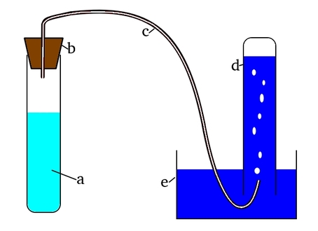
The setup includes a container a holding a 3% hydrogen peroxide H2O2 solution and catalase, sealed with a stopper b into which a flexible tube c is inserted. The other end of the tube leads into an inverted test tube d submerged in a vessel e filled with water. Initially, the test tube is completely filled with water, which is gradually displaced by the gas produced in the reaction.
One issue when using flexible tubing is keeping it bent in a shape that allows easy placement into the test tube. A simple solution is to insert a stiff wire, such as copper wire, about 1.5–2 mm (0.06–0.08 in) in diameter, as shown in Photo 3.

When inserted, the wire doesn’t block the tube but allows it to be shaped as needed (Photo 4).

The reaction vessel (Fig. 1a) can conveniently be replaced with a 10 cm3 (0.34 fl oz) syringe. The complete experimental setup is shown in Photo 5.

Approximately 4 cm3 (0.14 fl oz) of hydrogen peroxide is added to the syringe, followed by 2 cm3 (0.07 fl oz) of freshly prepared potato extract. Almost immediately, the reaction begins: the mixture foams vigorously and releases a large volume of gas (Photo 6).

The gas passes through the tube into the inverted test tube, displacing the water (Photo 7).

The gas produced is colorless and odorless: what exactly is it? To identify it, you can insert a glowing splint into the test tube. Alternatively, bring the gas outlet close to a glowing splint (Photo 8A). In both cases, the splint reignites with a bright flame (Photo 8B).

This test confirms that the gas produced is oxygen O2, which enables combustion. In an oxygen-rich environment or in pure oxygen, combustion occurs much more vigorously.
For the next experiment, we’ll use a different source: baker’s yeast Saccharomyces cerevisiae, particularly fresh or compressed (Photo 9).

Yeast, which are unicellular fungi, are one of the most widely used microorganisms by humans.
To prepare a yeast suspension, mix a small amount of yeast with lukewarm water (around 95°F or 35°C). The resulting suspension contains dispersed yeast cells (Photo 10).

Into three test tubes, add a few cm3 of hydrogen peroxide mixed with dish soap (Photo 11A). Tube a serves as the control and contains 1 cm3 (0.03 fl oz) of distilled water. Tube b contains the same amount of yeast suspension, while tube c contains yeast suspension that has been briefly boiled and cooled to room temperature.

Within seconds, noticeable changes appear (Photo 11B). Tube b begins to foam; evidence that yeast cells are rich in catalase. No such effect is observed in tube c, where the yeast has been heat-treated.
As a side note, the test tube rack shown in the photo was constructed from syringe pieces glued to a plastic base. This design provides greater visibility from all sides, is inexpensive, and is simple to build in a school.
Returning to the experiment: the foam forms because the oxygen is trapped. Even with small liquid volumes, a substantial amount of stable foam is produced (Photo 12).

The lack of reaction in control tube a is expected. But why is there no foaming in tube c, even though it contains yeast?
Explanation
Catalase promotes the decomposition of hydrogen peroxide into water and oxygen, as shown in the equation:
Hydrogen peroxide is unstable and spontaneously decomposes. Under normal conditions, especially in dilute solutions, this reaction is relatively slow. The addition of catalase drastically accelerates the reaction, as we saw in our experiments.
We often overlook the fact that living in an oxygen-rich environment is a considerable challenge for organisms. Although aerobic respiration yields more energy than anaerobic processes, it also leads to the formation of reactive oxygen species (ROS), which can damage cellular components. Hydrogen peroxide is one such ROS. It forms as a by-product of various metabolic pathways and must be quickly converted to less reactive substances to protect the organism. Molecular oxygen is far less reactive than peroxides, so catalase plays a protective role in preventing cellular damage [5].
From this perspective, oxygen, at least in some forms, can be considered toxic. Organisms that utilize it for respiration have evolved defense mechanisms, including enzymes like catalase. It’s no surprise that catalase is found in nearly all organisms living in oxygen-rich environments. It’s also one of the most efficient enzymes known, capable of catalyzing millions of hydrogen peroxide breakdown reactions per second [6].
The catalase molecule has a tetrameric structure, composed of four polypeptide chains (each over 500 amino acids). As a protein, it is susceptible to denaturation, meaning that high temperatures irreversibly damage its structure and activity, as observed in test tube c of the second experiment.
Catalase activity can also be inhibited by heavy metal ions. Human catalase functions optimally near neutral pH. Exploring how different factors influence the catalytic activity of this enzyme could be the focus of further simple yet insightful experiments.
References:
- [1] Berg J. M., Tymoczko J. L., Stryer L., Biochemia, Wydawnictwo Naukowe PWN, Warszawa, 2007 back
- [2] Mayes P. A., Bioenergetyka: rola ATP, in: Murray R. K., Kokot F., Koj A., Aleksandrowicz Z., Biochemia Harpera, Wydawnictwo Lekarskie PZWL, Warszawa, 2006, pp. 159-166 back
- [3] Ples M., Enzymy - biologiczne katalizatory (eng. Enzymes: Catalysts of Life), Chemia w Szkole (eng. Chemistry in School), 3 (2016), Agencja AS Józef Szewczyk, pp. 6-11 back
- [4] Sumner J. B., Dounce A. L., Crystalline Catalase, Science, 85 (2206), 1937, pp. 366-367 back
- [5] Gaetani G. F., Ferraris A. M., Rolfo M., Mangerini R., Arena S., Kirkman H. N., Predominant role of catalase in the disposal of hydrogen peroxide within human erythrocytes, Blood, 87 (4), 1996, pp. 1595-1599 back
- [6] Goodsell D. S., Catalase, online: http://pdb101.rcsb.org/motm/57 [28.11.2018] back
All photographs and illustrations were created by the author.
Marek Ples